Madeline’s Story

Site created on February 16, 2019

Madeline was diagnosed with Neuroblastoma in February 2019 at age 3. Sedona was diagnosed in June 2019 at age one with Isolated Hemihypertrophy which puts her at higher risk for developing childhood cancer. This is our story.

How Can You Help?
- Meal Train - https://www.mealtrain.com/trains/q3yo81
- Donate Blood - Madeline is type A+ (she can accept 0+/- and A+/- but please donate no matter your type if you can!
- If you live far away and still want to help we would love some assistance with extra child care expenses. Please venmo @bethany-cermak
- Send a card or bead of support
- Send a cleaning crew to our house
- Donate to a charity in honor of Madeline

Please note: If you donate through this site, you are supporting caringbridge.org and not us directly.

Newest Update

Journal entry by Bethany Cermak

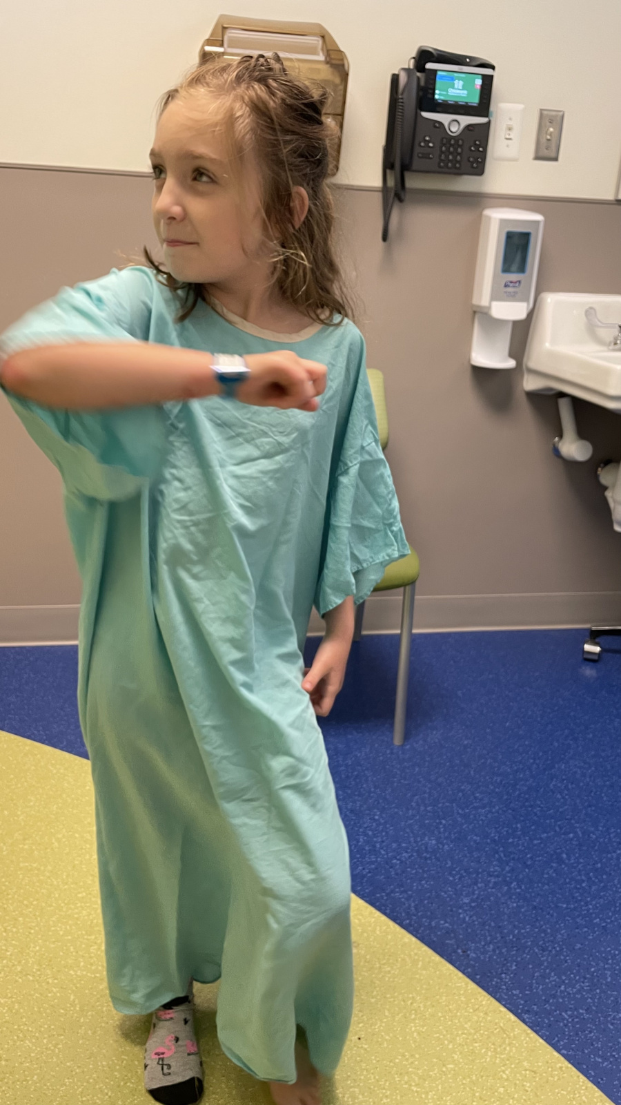
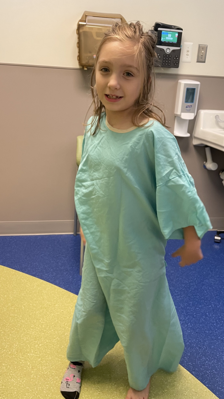
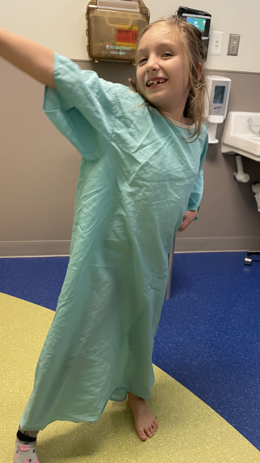
Madeline had an MRI yesterday where she was awarded star patient of the week! It was not a real prize and it's entirely possible they tell everyone they've earned it but she really was such a rockstar! It's been a while since she's had scans so she didn't remember the part where she gets an IV and she freaked out for roughly 2 minutes, pulled herself together, and let them do it. She watched the lego movie through a series of mirrors and stayed completely still while getting her MRI. Her bravery and maturity always amazes me!

We're going to meet with ENT and Audiology over the next few weeks and then have her survivorship appointment November 1st!

Full MRI Report for those who are interested:

EXAMINATION: MRI ABD MASS (ABD&PEL, W/WO)
 
INDICATION: 7-year-old female with intermediate-risk neuroblastoma; disease surveillance. Large right-sided pelvic mass with L3-sacral intraspinal extension, status post partial tumor resection and chemotherapy, persistent elevated HVA/VMA.
 
TECHNIQUE: Multisequence, multiplanar MR imaging of the abdomen and pelvis was performed according to standard departmental protocol on a 3Tesla magnet. Imaging was acquired prior to and following the administration of 2.7 mL of Gadavist intravenously.
 
COMPARISON: MR of the abdomen and pelvis dated January 19, 2023

IMPRESSION:    
1. Unchanged size and extent of lumbosacral right greater than left paraspinal mass with intraspinal extension, involvement of the right greater than left psoas and paraspinal muscles, and extension along the right lumbosacral plexus/sciatic nerve.
2. No new abdominal pelvic adenopathy or more distant metastases.
 
FINDINGS: 
 
ABDOMEN:
Liver: Hepatic signal is normal with no focal lesions. No evidence of liver fat or iron deposition. After contrast administration, no enhancing lesions are identified.
 
Bile ducts/Gallbladder: No intra or extrahepatic biliary ductal dilatation. No biliary ductal filling defects. The gallbladder is normal with no wall thickening, pericholecystic fluid, or gallstones.
 
Pancreas: Normal signal throughout with no focal lesions. No pancreatic ductal dilatation. Normal enhancement after contrast administration.
 
Spleen: Normal in size and appearance.
 
Kidneys/Adrenals: Both kidneys are normal in size and appearance. No urinary tract dilatation. Unchanged simple renal cyst in the superior pole of the left kidney. Enhancement and excretion is symmetric. Normal adrenal glands.
 
ABDOMEN/PELVIS:
Bowel: Normal bowel loops throughout the abdomen with no wall thickening or dilation.
 
Vessels: Normal enhancement of the hepatic artery, portal vein, hepatic veins, superior mesenteric vein, and splenic vasculature. The aorta and inferior vena cava are patent. Incidentally seen is a replaced left hepatic artery.
 
Peritoneum: No ascites.
 
Bones: Unchanged T2-hyperintense focus in the center of the L5 vertebra and mild height loss at L4. Background marrow signal within normal limits.
 
PELVIS:
General: Allowing for differences in technique, similar appearance of mildly enhancing, T2 hyperintense mass located at the lumbosacral junction mostly in the right pelvis but extending across midline/into the spinal canal at multiple neural foramen into the left pelvis. The intraspinal component of the mass demonstrates unchanged compression of thecal sac. L4-S1 with unchanged expansion of the bilateral L4-L5 and L5-S1 neural foramen and contouring of the posterior L4- S1 vertebral bodies. 
 
The right pelvic/paraspinal component measures approximately 5.6 x 5 cm (AP by transverse, series 905 image 102), previously 5.2 x 5.3 cm. There is unchanged extension/involvement of the mass into the right paraspinal musculature and right psoas with extension of the mass along the right lumbosacral plexus and sciatic nerve. The mass crosses midline and involves the left psoas, although to a lesser extent in the right psoas. The left paraspinal component is measured at 3.1 x 1.9 cm, previously 2.8 x 1.9 cm, stable underlying for measurement differences. The extent of the mass is similar to prior exam. Multiple foci of susceptibility artifact in the midline lower abdomen/pelvis in the region of the mass in keeping with multiple surgical clips.
 
Uterus/ovaries: No uterine abnormality. Ovaries not well seen in keeping with prepubertal status.
 
Bladder: Normal.
 
The right common iliac vein is not well visualized.
Patients and caregivers love hearing from you; add a comment to show your support.
Help Madeline Stay Connected to Family and Friends

A $25 donation to CaringBridge powers a site like Madeline's for two weeks. Will you make a gift to help ensure that this site stays online for them and for you?

Comments Hide comments

Show Your Support

See the Ways to Help page to get even more involved.

SVG_Icons_Back_To_Top
Top