Welcome to Doug’s CaringBridge Site
Sign In to Show Your Support
My results from the MRI and Dr. visit are as follows:
FINDINGS: There are postoperative changes of left-sided craniotomy. There is hemosiderin staining deep to the craniotomy. Small nodular and curvilinear enhancing foci. The craniotomy is similar to prior when allowing for differences in technique. There is no new masslike enhancement at this site. Again noted are several enhancing intracranial lesions, as detailed below: * There has been continued increase in size of an enhancing intra-axial lesion in the right occipital lobe that measures 1.7 x 1.3 x 1.4 cm (AP x TV x CC) and previously measured 1.0 x 0.9 x 0.9 cm on the 1/30/2024 exam. There has been accompanying increase in vasogenic edema in the right occipital lobe and periatrial region surrounding the lesion. MR perfusion imaging is nondiagnostic. * A 5 x 6 x 7 mm enhancing nodule in the parasagittal right frontal lobe is similar to very minimally increased in size from the most recent exam and certainly increased in size from the 10/9/2023 study (series 16, image 133). A heterogeneous focus of enhancement just posterior to the above lesion and abutting the falx has not significantly changed from the prior exam.
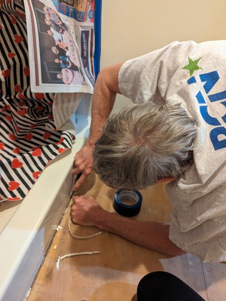
As I understand it, the lesions are increasing in size so we have to do something. The 1st choice is that I have already started steroids which helped before. The benefit is more energy to get work done which why I have been digging dirt and rebuilding a retaining wall under the pool for the last 3 days. I am thankful it is raining and wet out today so I can rest my knees and back. I also have an increase in my appetite but I am supposed to stay away from sugar. That just doesn't seem fair. The down sides are I have trouble sleeping and supposedly I am prone to being more irritable. I say just be nice to me, but Francine still thinks it is my fault for discussing any shortcomings I notice. Go figure. So If I happen to be short with any of you, please just blame the medication. This treatment will last for about 30 days and then off for another MRI to see if it helped. If not the next step is to drill probes into my head and try to zap the little buggers. Notice my expert use of medical terminology. Or they will open my head again and surgically remove the bad stuff and hopefully not take out any extra I need to be the kind, enjoyable, helpful, loving guy I am. As long as I am ready for camping and fun stuff in June.
As always, the Lord's will be done and we will trust in Him to know that all things work for the good of His people as He prepares us for our place in heaven to suit His purposes.
Thank you all for your love, concern, and prayers,
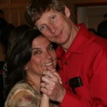
Doug and Francine Boone
8 Hearts • 3 Comments